ALABAMA: Tuberculosis and COVID-19 lung lesions revealed by high-resolution three-dimensional imaging
Researchers with the University of Alabama at Birmingham are using micro computer tomography, or microCT, to create a high resolution 3D atlas of lungs damaged by tuberculosis or COVID-19. “This atlas could be used to identify novel imaging biomarkers,” said senior author Adrie Steyn. Partial support for this research came from the NIH.
The research, published in the journal EMBO Molecular Medicine — with one of the 3D images featured on the journal cover, was led by Adrie Steyn, Ph.D., a professor in the Department of Microbiology at the University of Alabama at Birmingham and member of the Africa Health Research Institute, or AHRI, University of KwaZulu-Natal, in Durban, South Africa, along with UAB and AHRI researchers and colleagues at various South African institutions.
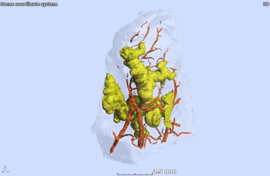
One of the benefits of microCT is creating a 3D image of a lesion. In a tuberculosis paper last year, Steyn and colleagues showed that power. For 70 years, they said, clinicians thought TB granulomas in the lungs of patients were spherical or ovoid because conventional histology showed round features, and researchers intuitively assumed those meant the granulomas were spherical or ovoid. But such round images are similar to cutting a very thin slice through a thick tree branch and assuming the branch is round or oval.
In Steyn’s current study, researchers also identified an unusual spatial arrangement of vasculature within an entire lobe of a COVID-19 lung, and 3D images of blood vessels revealed microangiopathy associated with hemorrhage.